
డయాలసిస్ చేయించుకునే ముందు తెలుసుకోవాల్సిన విషయాలు: రోగులు, కుటుంబ సభ్యులకు పూర్తి మార్గదర్శిని

డయాలసిస్ చేయించుకునే ముందు తెలుసుకోవాల్సిన విషయాలు: రోగులు, కుటుంబ సభ్యులకు పూర్తి మార్గదర్శిని కిడ్నీలు మన శరీరంలోని వ్యర్థ పదార్థాలను తొలగించడం, అదనపు నీటిని బయటకు పంపించడం, రక్తపోటును నియంత్రించడంలో ముఖ్యమైన పాత్ర పోషిస్తాయి. కిడ్నీల పనితీరు తీవ్రంగా తగ్గినప్పుడు శరీరంలో వ్యర్థ పదార్థాలు, ద్రవాలు పేరుకుపోవచ్చు. ఇలాంటి పరిస్థితుల్లో వైద్యులు రోగి ఆరోగ్యాన్ని బట్టి డయాలసిస్ (Dialysis) చికిత్సను సూచించవచ్చు. డయాలసిస్ ప్రారంభించాలని వైద్యులు చెప్పినప్పుడు రోగులకు, కుటుంబ సభ్యులకు అనేక సందేహాలు రావడం సహజం. చికిత్సకు ఎలా సిద్ధం కావాలి? ప్రతి సెషన్‌కు వెళ్లేటప్పుడు ఏం తీసుకెళ్లాలి? డయాలసిస్ యాక్సెస్‌ను ఎలా సంరక్షించాలి? చికిత్స తీసుకుంటూనే రోజువారీ పనులు చేసుకోవచ్చా? ఈ ప్రశ్నలకు సమాధానాలు తెలుసుకోవడం ద్వారా డయాలసిస్ ప్రయాణాన్ని మరింత సులభంగా నిర్వహించుకోవచ్చు. విజయవాడలో కిడ్నీ చికిత్స కోసం సరైన మార్గదర్శకత్వం కోరుకునే వారు American Kidney Institute, భారతి నగర్‌లో నెఫ్రాలజీ వైద్యుల సలహా తీసుకోవచ్చు. డయాలసిస్ అంటే ఏమిటి? డయాలసిస్ అనేది కిడ్నీలు తగినంతగా పనిచేయనప్పుడు రక్తంలోని వ్యర్థ పదార్థాలను, అదనపు ద్రవాలను తొలగించడంలో సహాయపడే చికిత్స. ఇది కిడ్నీల సహజ పనుల్లో కొన్నింటిని భర్తీ చేస్తుంది. డయాలసిస్‌లో ప్రధానంగా రెండు రకాలు ఉన్నాయి: హీమోడయాలసిస్ (Hemodialysis) ఈ విధానంలో రక్తాన్ని శరీరం నుంచి ప్రత్యేక ఫిల్టర్ ఉన్న డయాలసిస్ యంత్రం ద్వారా పంపిస్తారు. ఫిల్టర్ రక్తంలోని వ్యర్థ పదార్థాలు, అదనపు ద్రవాలను తొలగించడంలో సహాయపడుతుంది. ఆ తర్వాత శుద్ధి చేసిన రక్తాన్ని తిరిగి శరీరంలోకి పంపిస్తారు. పెరిటోనియల్ డయాలసిస్ (Peritoneal Dialysis) ఈ విధానంలో పొత్తికడుపులోని పెరిటోనియం అనే పొరను సహజ ఫిల్టర్‌గా ఉపయోగిస్తారు. ప్రత్యేక డయాలసిస్ ద్రవం సహాయంతో వ్యర్థ పదార్థాలు తొలగించబడతాయి. ఈ విధానం కొంతమంది రోగులకు అనుకూలంగా ఉంటుంది. అయితే దీనికి అవసరమైన శిక్షణ, పరిశుభ్రత, వైద్య పర్యవేక్షణ తప్పనిసరి. ఏ విధమైన డయాలసిస్ అనుకూలమో రోగి కిడ్నీ పనితీరు, ఇతర ఆరోగ్య సమస్యలు, జీవనశైలి, చికిత్స అవసరాల ఆధారంగా నెఫ్రాలజిస్ట్ నిర్ణయిస్తారు. డయాలసిస్ ప్రారంభించే ముందు ఎలా సిద్ధం కావాలి? డయాలసిస్ ప్రారంభించే ముందు వైద్యుడితో సరైన ప్రణాళిక రూపొందించుకోవడం ముఖ్యం. చికిత్సకు ముందస్తు సిద్ధత వల్ల అనవసరమైన ఇబ్బందులను తగ్గించుకోవచ్చు. 1. మీ చికిత్స ప్రణాళికను అర్థం చేసుకోండి డయాలసిస్ ఎన్ని సార్లు చేయించుకోవాలి, ప్రతి సెషన్ ఎంతసేపు ఉంటుంది, ఏ రకమైన డయాలసిస్ అవసరం వంటి విషయాలను వైద్యుడిని అడిగి తెలుసుకోండి. రోగులందరికీ ఒకే చికిత్స షెడ్యూల్ ఉండదు. 2. అవసరమైన పరీక్షల రిపోర్టులను సిద్ధంగా ఉంచుకోండి కిడ్నీ పనితీరుకు సంబంధించిన రక్త పరీక్షలు, ఎలక్ట్రోలైట్ పరీక్షలు, ఇతర వైద్య నివేదికలు, ప్రస్తుతం ఉపయోగిస్తున్న మందుల వివరాలను భద్రపరచండి. ప్రతి వైద్య సందర్శనలో ఈ వివరాలు ఉపయోగపడతాయి. 3. డయాలసిస్ యాక్సెస్ గురించి తెలుసుకోండి హీమోడయాలసిస్ కోసం రక్తాన్ని బయటకు తీసుకెళ్లి తిరిగి శరీరంలోకి పంపేందుకు ప్రత్యేక యాక్సెస్ అవసరం. దీనికి AV ఫిస్టులా, గ్రాఫ్ట్ లేదా కాథెటర్ వంటి విధానాలను ఉపయోగించవచ్చు. AV ఫిస్టులా చేయాల్సి ఉంటే, దాని కోసం సరైన సమయంలో ప్రణాళిక రూపొందించుకోవడం ముఖ్యం. ఏ యాక్సెస్ అనుకూలమో వైద్యుడు నిర్ణయిస్తారు. 4. ఆహారం, నీటి పరిమితుల గురించి అడగండి డయాలసిస్ రోగులందరికీ ఒకే రకమైన ఆహార నియమాలు ఉండవు. రక్త పరీక్షల ఫలితాలు, మూత్రం ఎంత వస్తోంది, శరీరంలో ద్రవాలు ఎంత పేరుకుపోతున్నాయి వంటి అంశాల ఆధారంగా ఆహారం, నీటి పరిమితులు మారుతాయి. మీకు ఎంత నీరు అవసరం, పొటాషియం లేదా ఫాస్ఫరస్ అధికంగా ఉన్న ఆహారాలను ఎంతవరకు పరిమితం చేయాలి అనే విషయాలను వైద్యుడు లేదా కిడ్నీ డైటీషియన్‌ను అడగండి. డయాలసిస్ సెషన్‌కు వెళ్లేటప్పుడు ఏం చేయాలి? ప్రతి డయాలసిస్ సెషన్‌కు ముందు కొన్ని సాధారణ జాగ్రత్తలు తీసుకోవడం ఉపయోగకరం. మీ మందులు, వైద్య నివేదికలు, చికిత్సకు సంబంధించిన వివరాలు వెంట తీసుకెళ్లండి. ఇంట్లో కొలిచిన రక్తపోటు వివరాలు ఉంటే వైద్య బృందానికి తెలియజేయండి. సెషన్‌కు ముందు శ్వాస తీసుకోవడంలో ఇబ్బంది, జ్వరం, ఛాతీ నొప్పి లేదా ఇతర కొత్త లక్షణాలు ఉంటే సిబ్బందికి చెప్పండి. డయాలసిస్ బృందం సూచించిన ఆహార నియమాలను పాటించండి. సెషన్‌ను స్వయంగా రద్దు చేయడం లేదా ఆలస్యం చేయడం కాకుండా, సమయానికి హాజరుకావడానికి ప్రయత్నించండి. డయాలసిస్‌కు ముందు ఎంత ఆహారం తీసుకోవాలి, మందులను ఎప్పుడు ఉపయోగించాలి అనే విషయాల్లో మీ చికిత్స బృందం సూచనలను అనుసరించండి. డయాలసిస్ యాక్సెస్‌ను ఎలా సంరక్షించాలి? హీమోడయాలసిస్ సురక్షితంగా జరగడానికి యాక్సెస్ సంరక్షణ చాలా ముఖ్యం. AV ఫిస్టులా లేదా గ్రాఫ్ట్ ఉన్నవారు: యాక్సెస్ ఉన్న చేతిపై అనవసరమైన ఒత్తిడి పడకుండా జాగ్రత్తపడండి. వైద్యుడు సూచించినట్లయితే ఫిస్టులాలో రక్త ప్రవాహం ఉందో లేదో క్రమం తప్పకుండా పరిశీలించండి. ఆ చేతిపై రక్తపోటు కొలవడం లేదా రక్తం తీసుకోవడం వంటి చర్యలకు ముందు వైద్య సిబ్బందికి ఫిస్టులా గురించి తెలియజేయండి. చేతిలో వాపు, ఎర్రదనం, నొప్పి, వేడి లేదా ఫిస్టులా స్పందనలో మార్పు కనిపిస్తే వెంటనే డయాలసిస్ బృందాన్ని సంప్రదించండి. కాథెటర్ ఉన్నవారు: కాథెటర్ ప్రాంతాన్ని శుభ్రంగా, పొడిగా ఉంచేందుకు వైద్య బృందం సూచనలు పాటించండి. కాథెటర్ డ్రెస్సింగ్‌ను మీరే తొలగించడం లేదా కనెక్షన్లను తెరవడం చేయకండి. జ్వరం, చలి, కాథెటర్ ప్రాంతంలో ఎర్రదనం, నొప్పి లేదా ద్రవం కారడం కనిపిస్తే వెంటనే వైద్య సహాయం పొందండి. యాక్సెస్‌ను సరిగ్గా సంరక్షించడం ఇన్ఫెక్షన్లు, చికిత్సకు అంతరాయాలు కలిగించే సమస్యల ప్రమాదాన్ని తగ్గించడంలో సహాయపడుతుంది. డయాలసిస్ తీసుకుంటూ సాధారణ జీవితాన్ని కొనసాగించవచ్చా? చాలా మంది రోగులు సరైన చికిత్స, ఆహార నియమాలు, వైద్య పర్యవేక్షణతో కుటుంబంతో సమయం గడపడం, తగిన పనులు చేయడం, తమ దైనందిన కార్యకలాపాలను కొనసాగించడం సాధ్యపడుతుంది. అయితే ప్రతి ఒక్కరి ఆరోగ్య పరిస్థితి వేర్వేరుగా ఉంటుంది. అలసట, రక్తహీనత, రక్తపోటు మార్పులు, ఇతర అనారోగ్య సమస్యలు రోజువారీ జీవితాన్ని ప్రభావితం చేయవచ్చు. జీవనశైలిని మెరుగ్గా నిర్వహించుకోవడానికి: వైద్యుడు సూచించిన డయాలసిస్ షెడ్యూల్‌ను పాటించండి. అనుమతించిన మేరకు తేలికపాటి శారీరక కార్యకలాపాలు చేయండి. తగినంత విశ్రాంతి తీసుకోండి. మందులను వైద్యుడి సూచనల ప్రకారం మాత్రమే వాడండి. కుటుంబ సభ్యులతో కలిసి చికిత్స షెడ్యూల్, ఆసుపత్రి సందర్శనలను ముందుగానే ప్రణాళిక చేసుకోండి. మానసిక ఒత్తిడి లేదా నిద్ర సమస్యలు ఉంటే వైద్యుడితో చర్చించండి. డయాలసిస్ తీసుకుంటున్నారని మాత్రమే అన్ని సాధారణ కార్యకలాపాలను పూర్తిగా మానేయాల్సిన అవసరం లేదు. మీ ఆరోగ్యానికి అనుగుణంగా ఏ పనులు చేయవచ్చో వైద్యుడి సలహా తీసుకోండి. డయాలసిస్ సమయంలో ఎప్పుడు వెంటనే వైద్య సహాయం తీసుకోవాలి? కొన్ని లక్షణాలు తీవ్రమైన సమస్యకు సంకేతాలు కావచ్చు. వాటిని నిర్లక్ష్యం చేయకూడదు. తీవ్రమైన శ్వాస తీసుకోవడంలో ఇబ్బంది ఛాతీ నొప్పి స్పృహ కోల్పోవడం లేదా తీవ్రమైన గందరగోళం ఆగని రక్తస్రావం జ్వరంతో పాటు చలి లేదా ఇన్ఫెక్షన్ అనుమానం ఫిస్టులా స్పందన అకస్మాత్తుగా లేకపోవడం లేదా యాక్సెస్‌లో తీవ్రమైన మార్పులు తీవ్రమైన లక్షణాలు ఉంటే వెంటనే అత్యవసర వైద్య సహాయం పొందండి. తదుపరి డయాలసిస్ సెషన్ వరకు వేచి ఉండకండి. విజయవాడలో డయాలసిస్ చికిత్స కోసం ఎక్కడ సంప్రదించాలి? డయాలసిస్ చికిత్స కోసం ఆసుపత్రిని ఎంపిక చేసేటప్పుడు చికిత్స అందుబాటు మాత్రమే కాకుండా, నెఫ్రాలజీ పర్యవేక్షణ, డయాలసిస్ యాక్సెస్ సంరక్షణ, రక్త పరీక్షల పర్యవేక్షణ, ఇన్ఫెక్షన్ నియంత్రణ, ఆహార మార్గదర్శకత్వం వంటి అంశాలను కూడా పరిగణనలోకి తీసుకోవాలి. American Kidney Institute, భారతి నగర్, విజయవాడలో కిడ్నీ సంబంధిత సమస్యల కోసం వైద్య మూల్యాంకనం, నెఫ్రాలజీ సంప్రదింపులు మరియు డయాలసిస్ చికిత్సకు సంబంధించిన మార్గదర్శకత్వం కోసం సంప్రదించవచ్చు. డయాలసిస్ అవసరం ఉందా, ఏ విధమైన చికిత్స అనుకూలం, ఎంత తరచుగా చికిత్స తీసుకోవాలి అనే నిర్ణయాలను రోగి ఆరోగ్య పరిస్థితిని పరిశీలించిన తర్వాత వైద్యులు తీసుకుంటారు. కిడ్నీ పనితీరు తగ్గినట్లు తెలిసిన వెంటనే సరైన వైద్య సలహా తీసుకోవడం, చికిత్స ప్రణాళికను అర్థం చేసుకోవడం, క్రమం తప్పకుండా పర్యవేక్షణ పొందడం రోగి ఆరోగ్య నిర్వహణలో ముఖ్యమైనవి. తరచుగా అడిగే ప్రశ్నలు (FAQs) 1. డయాలసిస్ ఎప్పుడు అవసరం అవుతుంది? కిడ్నీలు శరీరంలోని వ్యర్థ పదార్థాలు, అదనపు ద్రవాలను తగినంతగా తొలగించలేని స్థితిలో డయాలసిస్ అవసరం కావచ్చు. ఈ నిర్ణయం కేవలం క్రియాటినిన్ స్థాయిపై కాకుండా లక్షణాలు, రక్త పరీక్షలు, ద్రవాల సమతుల్యత, మొత్తం ఆరోగ్య పరిస్థితి ఆధారంగా తీసుకుంటారు. 2. డయాలసిస్ ప్రారంభించే ముందు ఫిస్టులా తప్పనిసరిగా చేయించుకోవాలా? ప్రతి రోగికి ఒకే విధమైన యాక్సెస్ అవసరం ఉండదు. దీర్ఘకాలిక హీమోడయాలసిస్ అవసరమయ్యే కొంతమందికి AV ఫిస్టులా అనుకూలంగా ఉంటుంది. ఇతర సందర్భాల్లో గ్రాఫ్ట్ లేదా కాథెటర్ అవసరమవచ్చు. మీకు ఏది సరైనదో వైద్యుడు నిర్ణయిస్తారు. 3. డయాలసిస్ సెషన్‌ను మిస్ అయితే ఏమవుతుంది? సెషన్ మిస్ కావడం వల్ల శరీరంలో వ్యర్థ పదార్థాలు, అదనపు ద్రవాలు లేదా పొటాషియం పేరుకుపోయే ప్రమాదం ఉంది. ఇది కొంతమందిలో తీవ్రమైన సమస్యలకు దారితీయవచ్చు. సెషన్‌కు హాజరుకాలేని పరిస్థితి వస్తే వెంటనే మీ డయాలసిస్ బృందాన్ని సంప్రదించండి. 4. డయాలసిస్ తీసుకునేవారు ఎంత నీరు తాగవచ్చు? అందరికీ ఒకే పరిమాణం సరిపోదు. మూత్రం ఎంత వస్తోంది, శరీరంలో వాపు ఉందా, గుండె పరిస్థితి, రక్త పరీక్షలు, డయాలసిస్ షెడ్యూల్ వంటి అంశాల ఆధారంగా నీటి పరిమితి నిర్ణయించబడుతుంది. మీ వైద్యుడు సూచించిన పరిమితిని పాటించండి. 5. డయాలసిస్ తీసుకునేవారు పని చేయవచ్చా? చాలా మంది రోగులు తమ ఆరోగ్య పరిస్థితికి అనుగుణంగా పని లేదా ఇతర రోజువారీ కార్యకలాపాలను కొనసాగించగలరు. అయితే పని సమయం, అలసట, చికిత్స షెడ్యూల్, ఇతర అనారోగ్య సమస్యలను పరిగణనలోకి తీసుకొని వైద్యుడితో ప్రణాళిక రూపొందించుకోవాలి. 6. విజయవాడలో డయాలసిస్ చికిత్స కోసం ఎవరిని సంప్రదించాలి? కిడ్నీ పనితీరు తగ్గినప్పుడు లేదా డయాలసిస్ అవసరం గురించి సందేహాలు ఉన్నప్పుడు
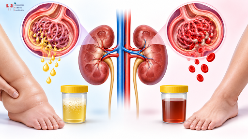

Understanding Nephrotic Syndrome and Nephritic Syndrome in Glomerulonephritis

Understanding Nephrotic Syndrome and Nephritic Syndrome in Glomerulonephritis The kidneys play an important role in maintaining the body’s overall health. They filter the blood, remove waste products, balance fluids and electrolytes, and help control blood pressure. Tiny structures called glomeruli perform much of this filtering work. When the glomeruli become inflamed or damaged, a condition called glomerulonephritis can develop. Depending on the type and severity of glomerular damage, a person may develop different patterns of kidney problems. Two important clinical patterns are nephrotic syndrome and nephritic syndrome. Although both can occur due to glomerular disease, they do not present in exactly the same way. Nephrotic syndrome is mainly associated with heavy protein loss through the urine, low blood albumin levels, and significant swelling. Nephritic syndrome is more commonly associated with blood in the urine, inflammation of the glomeruli, reduced kidney function, and high blood pressure. Understanding the difference between these two patterns can help people recognize warning signs and seek appropriate medical evaluation at the right time. What Is Glomerulonephritis? Glomerulonephritis refers to inflammation or damage involving the glomeruli, which are tiny blood-filtering units inside the kidneys. Healthy glomeruli allow water and small waste products to pass into the urine while keeping important substances such as blood cells and most proteins inside the bloodstream. When the glomerular filtering barrier becomes damaged, its normal function can change. Protein may start leaking into the urine, blood cells may enter the urine, and the kidneys may become less effective at removing waste. The clinical symptoms depend on the specific type of glomerular disease. Some people may have mild changes detected only through urine or blood tests. Others may develop noticeable swelling, dark or reddish urine, reduced urine output, or increased blood pressure. Glomerulonephritis can occur suddenly or develop gradually. Some forms are temporary, while others can progress and cause chronic kidney disease if the underlying cause is not identified and managed appropriately. What Is Nephrotic Syndrome? Nephrotic syndrome is a group of symptoms and laboratory findings that occur when the glomeruli allow an unusually large amount of protein to pass into the urine. One of the most important features is heavy proteinuria. Proteinuria means the presence of excess protein in the urine. Albumin is an important protein in the blood. It helps maintain the normal movement of fluid between blood vessels and body tissues. When large quantities of albumin are lost through the urine, the blood albumin level can fall. This condition is known as hypoalbuminemia. As albumin decreases and the body’s fluid balance changes, fluid can move into surrounding tissues. This can result in swelling called edema. In severe cases, swelling can become widespread throughout the body. This severe generalized swelling may be described as anasarca. Common Features of Nephrotic Syndrome The main features may include: Heavy protein loss in urine: Large amounts of protein can pass through damaged glomeruli, resulting in significant proteinuria that can be detected through urine testing. Foamy or frothy urine: Excess protein in urine may make the urine appear unusually foamy, although foamy urine can also occur for other reasons. Low blood albumin: Continuous urinary protein loss can reduce the albumin concentration in the bloodstream. Swelling of the body: Edema may first become noticeable around the eyes, feet, ankles, or legs and may become more widespread. Weight gain from fluid retention: A sudden increase in body weight can sometimes result from accumulation of excess fluid rather than an increase in body fat. Increased risk of infections and blood clots: Some forms of nephrotic syndrome can increase the risk of complications, making medical monitoring important. Why Does Nephrotic Syndrome Cause Swelling? Swelling is one of the most noticeable symptoms of nephrotic syndrome. The kidneys may lose large amounts of protein through the urine. Albumin is particularly important because it helps maintain oncotic pressure within blood vessels. When blood albumin becomes significantly low, fluid can move more easily from blood vessels into surrounding tissues. The kidneys may also retain more sodium and water as the body attempts to restore circulating fluid volume. This can further increase swelling. The result may be edema in the legs, feet, face, abdomen, or other parts of the body. In severe cases, fluid accumulation may become widespread and cause anasarca. What Is Nephritic Syndrome? Nephritic syndrome is another clinical pattern associated with glomerular inflammation and damage. Unlike nephrotic syndrome, where heavy protein loss is the major feature, nephritic syndrome is commonly characterized by hematuria, reduced kidney filtration, and increased blood pressure. Hematuria means blood is present in the urine. When red blood cells pass through inflamed glomeruli, the urine may become pink, red, brown, or cola-colored. However, blood in urine is not always visible to the naked eye. Sometimes it can only be detected through urine microscopy or other laboratory testing. Inflammation within the glomeruli can also reduce the kidney’s ability to filter blood efficiently. As kidney filtration decreases, waste products can accumulate in the bloodstream. Fluid and sodium retention may also contribute to high blood pressure. Common Features of Nephritic Syndrome The symptoms and findings may include: Hematuria: Blood may leak through damaged glomeruli and appear in the urine as microscopic or visible blood. Dark or cola-colored urine: Significant blood in the urine can change its appearance, although urine color alone cannot confirm the cause. High blood pressure: Reduced kidney filtration and fluid retention can contribute to elevated blood pressure. Reduced urine output: Some people may produce less urine when kidney filtration is significantly affected. Mild to moderate edema: Swelling can occur, but it is often less prominent than the widespread edema seen in severe nephrotic syndrome. Reduced kidney function: Blood tests may show an increase in creatinine when the kidneys are not filtering normally. Nephrotic Syndrome vs Nephritic Syndrome Although both conditions involve the glomeruli, their typical clinical patterns are different. Nephrotic Syndrome The dominant problem is heavy protein leakage. Patients may notice: Foamy urine Significant proteinuria Low albumin levels Swelling around the eyes Swelling of the legs and feet Generalized

Which Is Better for Dialysis: Catheter or AV Fistula? A Complete Guide

Which Is Better for Dialysis: Catheter or AV Fistula? A Complete Guide Starting hemodialysis is a major change in a patient’s life. Along with understanding the dialysis process, one of the most important decisions is choosing the right vascular access. Vascular access is the route through which blood leaves the body, travels to the dialysis machine for cleaning, and returns to the bloodstream. Without reliable vascular access, hemodialysis cannot be performed effectively. The two options patients commonly hear about are a dialysis catheter and an AV fistula. Although both can provide access for hemodialysis, they are very different in how they are created, how quickly they can be used, and the risks associated with them. For many patients who are expected to need long-term hemodialysis, an AV fistula is generally preferred because it usually has fewer complications and better long-term durability than a central venous catheter. However, a catheter can be extremely useful when dialysis needs to begin urgently or when a fistula or graft is not suitable. For patients looking for a Kidney Specialist in Vijayawada, understanding these differences can make conversations about dialysis planning much easier. What Is Vascular Access for Hemodialysis? During hemodialysis, blood needs to move from the patient’s body to a dialysis machine. The machine passes the blood through a special filter called a dialyzer, where waste products and excess fluid are removed. The cleaned blood is then returned to the body. A vascular access provides this connection between the patient’s bloodstream and the dialysis machine. There are three major types of hemodialysis vascular access: AV fistula: A surgeon connects an artery and a nearby vein, usually in the arm, creating a stronger blood vessel that can later be used for dialysis needles. AV graft: A special tube is used to connect an artery and vein when the patient’s own blood vessels are not suitable for a fistula. Dialysis catheter: A soft tube is placed into a large vein, commonly in the neck or chest, allowing blood to be taken from and returned to the body without placing needles into an AV access. Each option has a role, but they are not interchangeable in every situation. What Is an AV Fistula? An arteriovenous fistula, commonly called an AV fistula or simply a fistula, is a surgically created connection between an artery and a nearby vein. It is usually created in the arm. The connection allows arterial blood flow to enter the vein, causing the vein to become larger and stronger over time. Once the fistula has matured sufficiently, dialysis needles can be placed into it during hemodialysis. An AV fistula uses the patient’s own blood vessels rather than an artificial tube. This is one of the major reasons it is generally preferred for long-term hemodialysis when it is medically suitable. The National Kidney Foundation describes a fistula as the preferred access because it generally has fewer problems and lasts longer than other access types. What Is a Dialysis Catheter? A dialysis catheter is a soft, flexible tube placed into a large vein. For hemodialysis, the catheter is commonly placed in a large vein in the neck, although other sites may be considered depending on the patient’s circumstances. Unlike a fistula, the catheter does not require the patient to wait for the access to mature before it can be used. This makes it particularly useful when dialysis needs to start urgently. For example, a patient who develops severe acute kidney injury and suddenly requires dialysis may not have enough time to wait for a fistula to mature. In such situations, a catheter may provide immediate access. However, because the catheter enters a large vein and has an external component, it carries higher risks of infection and other access-related complications than a well-functioning AV fistula. AV Fistula vs Dialysis Catheter: Which Is Better? For a patient who needs long-term hemodialysis and has suitable blood vessels, an AV fistula is generally preferred over a dialysis catheter. This does not mean that a catheter is a “bad” option. A catheter can be the right option in an emergency, during the period before a fistula is ready, or when an AV fistula or graft cannot be created. The difference is mainly about the intended duration, patient’s condition, vascular anatomy, and urgency of dialysis. A fistula generally offers better long-term durability and a lower risk of infection than a catheter. A catheter, on the other hand, provides rapid access and does not require maturation. Why Is an AV Fistula Preferred for Long-Term Dialysis? There are several reasons why an AV fistula is generally preferred when it is appropriate for the patient. Lower Infection Risk One of the major advantages of a fistula is the lower risk of bloodstream infection compared with a dialysis catheter. A catheter creates a direct pathway through the skin into a large vein. Because part of the catheter remains outside the body, strict infection-control practices are essential. A mature fistula is completely under the skin between dialysis sessions. This does not mean that a fistula can never become infected, but its infection risk is generally lower than that of a central venous catheter. Better Long-Term Durability An appropriately functioning AV fistula can provide vascular access for a long period. Because it is created using the patient’s own blood vessels, it does not depend on a synthetic tube or an external catheter. However, fistulas can develop problems such as narrowing, clotting, aneurysmal changes, or failure to mature. Regular monitoring helps identify these issues early. Good Blood Flow A mature fistula is designed to provide the blood flow needed for hemodialysis. During dialysis, two needles are typically placed into the fistula. One needle allows blood to travel toward the dialysis machine, while the other returns the cleaned blood to the patient. Good blood flow is important for delivering an effective dialysis treatment. Why Is a Dialysis Catheter Used? Although fistulas are generally preferred for long-term access, dialysis catheters remain very important. The biggest advantage is immediate usability. A

Does Starting Dialysis Mean You Need It for Life? AKI vs CKD Explained

Does Starting Dialysis Mean You Need It for Life? AKI vs CKD Explained Starting dialysis can be frightening for both patients and their families. One of the first questions many people ask is, “Once dialysis starts, will I have to continue it for the rest of my life?” The answer is not always. Whether dialysis is temporary or long-term depends mainly on why the kidneys stopped working and whether the kidney damage can recover. Acute Kidney Injury (AKI) and Chronic Kidney Disease (CKD) are two very different conditions, and the need for dialysis can be very different in each. In some patients with AKI, dialysis may be needed only for a few days or weeks while the kidneys recover. In advanced CKD, however, the kidneys may have permanent damage, and long-term dialysis or kidney transplantation may become necessary. For anyone searching for the Best Nephrologist in Vijayawada, understanding the difference between AKI and CKD is an important first step in understanding why dialysis has been recommended. What Is Dialysis? Dialysis is a treatment that performs some of the important functions normally carried out by healthy kidneys. Healthy kidneys remove waste products and excess fluid from the blood. They also help maintain the body’s electrolyte and acid-base balance. When kidney function becomes severely reduced, waste products, excess fluid, potassium, and acids can build up in the body. Dialysis helps remove these substances when the kidneys are unable to do so adequately. There are two main types of dialysis: Hemodialysis: Blood is passed through a special filter called a dialyzer, where waste products and excess fluid are removed before the blood is returned to the body. Peritoneal dialysis: The patient’s peritoneum, which is the lining inside the abdomen, is used as a natural filtering membrane with the help of dialysis fluid. The type of dialysis depends on the patient’s medical condition, kidney function, lifestyle, treatment needs, and medical advice. When patients search for Dialysis in Vijayawada, they should understand that dialysis is not a single treatment plan that is identical for every patient. The duration and frequency depend on the underlying kidney condition. Does Starting Dialysis Mean It Will Continue for Life? No. Starting dialysis does not automatically mean that dialysis will be lifelong. This is one of the most important things patients should understand. The kidneys can sometimes recover after a sudden injury. If dialysis was started because of an episode of AKI, the treatment may be temporary. For example, severe infection, dehydration, very low blood pressure, certain medicines, or other medical problems can suddenly affect kidney function. If the underlying problem is treated and the kidneys recover, dialysis may eventually be stopped. On the other hand, advanced CKD usually develops over many years. When a large amount of kidney tissue has been permanently damaged, the kidneys may not recover enough to perform their essential functions independently. In such situations, long-term dialysis or kidney transplantation may be required. Therefore, the most important question is not simply “Have I started dialysis?” The more important question is: “Why did my kidneys stop working?” What Is Acute Kidney Injury? Acute Kidney Injury, commonly called AKI, is a sudden decrease in kidney function. It can develop within hours or days. Unlike CKD, AKI may be reversible, depending on the cause and severity of the injury. Some patients with severe AKI may temporarily need dialysis because their kidneys cannot remove enough waste and fluid from the body. However, once the underlying cause is treated and kidney function improves, dialysis may no longer be necessary. This is why patients with AKI should not immediately assume that dialysis will continue permanently. Common Causes of Acute Kidney Injury AKI can occur for many different reasons. Some of the common causes include: Severe infections such as sepsis: A serious infection can cause low blood pressure, inflammation, and reduced blood flow to the kidneys, leading to sudden kidney injury. Severe dehydration: Significant loss of fluids due to vomiting, diarrhea, excessive sweating, poor fluid intake, or other causes can reduce blood flow to the kidneys. Very low blood pressure: Conditions such as severe infection, major blood loss, or shock can reduce the blood supply reaching the kidneys. Certain medicines or toxins: Some medicines can injure the kidneys, especially when used inappropriately or in vulnerable patients. Urinary obstruction: Problems that block the normal flow of urine can sometimes cause kidney function to deteriorate. Inflammatory or immune-related kidney diseases: Certain conditions can directly affect kidney tissue and cause sudden deterioration in kidney function. The treatment of AKI focuses on identifying and correcting the underlying cause while supporting the patient until the kidneys recover. When Is Dialysis Needed in AKI? Not every patient with AKI requires dialysis. Doctors decide whether dialysis is necessary based on the patient’s overall condition rather than one blood test alone. Dialysis may be considered when kidney failure leads to problems that cannot be safely managed with medicines and supportive treatment. Some important situations include: Severe fluid overload: When the kidneys cannot remove excess fluid and the patient develops serious fluid accumulation, dialysis may be needed to remove the extra fluid. Dangerously high potassium levels: Severe hyperkalemia can affect the heart and may require urgent treatment, including dialysis in selected situations. Severe metabolic acidosis: When the body becomes excessively acidic and the problem cannot be adequately corrected with medical treatment, dialysis may be considered. Significant accumulation of waste products: Severe buildup of waste products can cause symptoms and complications that may require dialysis. Certain severe complications of kidney failure: The patient’s symptoms and overall clinical condition may make dialysis necessary. In these situations, dialysis acts as supportive treatment while doctors work to treat the cause of AKI. Can Dialysis Be Stopped After AKI? Yes, dialysis can sometimes be stopped when kidney function recovers sufficiently. This is one of the biggest differences between temporary dialysis for AKI and maintenance dialysis for advanced CKD. When a patient is receiving dialysis for AKI, the nephrology team regularly monitors kidney function and the patient’s

Kidney Biopsy for Glomerulonephritis, AKI, CKD, and Transplant Rejection

Kidney Biopsy for Glomerulonephritis, AKI, CKD, and Transplant Rejection Kidney diseases do not always show their exact cause through routine blood and urine tests. Tests such as serum creatinine, eGFR, urine protein, and urine analysis can tell us that the kidneys are not functioning normally. However, in some patients, these tests cannot tell us exactly what is happening inside the kidney. This is where a kidney biopsy can become important. A kidney biopsy is a diagnostic procedure in which a very small piece of kidney tissue is removed and examined under a microscope. The tissue can provide detailed information about the type of kidney disease, the pattern of kidney injury, inflammation, scarring, and other changes that may not be visible through routine tests. A kidney biopsy may be recommended in conditions such as glomerulonephritis, unexplained acute kidney injury (AKI), chronic kidney disease (CKD) of unknown cause, significant protein in the urine, and suspected rejection after a kidney transplant. The information obtained from a biopsy can help the nephrologist understand the disease more accurately and select an appropriate treatment plan. For patients looking for specialized kidney care, choosing a Best Kidney Hospital in Vijayawada can provide access to nephrology evaluation, diagnostic testing, biopsy services, and appropriate follow-up care under specialist supervision. What Is a Kidney Biopsy? A kidney biopsy is a procedure used to obtain a small sample of kidney tissue for detailed laboratory examination. Although blood and urine tests are extremely useful, they mainly provide information about kidney function and the substances being removed or retained by the kidneys. A biopsy can provide a much closer look at the kidney tissue itself. The sample is usually examined using different laboratory techniques. These may include routine microscopic examination, immunological staining, and electron microscopy when required. These examinations can help identify specific patterns of kidney injury. For example, two patients may both have high serum creatinine and protein in their urine, but the underlying diseases may be completely different. One patient may have an immune-related kidney disease, while another may have a different type of glomerular injury. The treatment for these conditions may also be different. Therefore, understanding the exact kidney disease can be important before starting certain treatments. Why Is a Kidney Biopsy Needed? A kidney biopsy is not required for every person with kidney disease. The decision is usually based on the patient’s symptoms, blood test results, urine findings, imaging results, medical history, and the suspected kidney condition. A nephrologist may recommend a biopsy when knowing the exact cause of kidney damage is likely to change the treatment or help determine the expected outcome. A biopsy may help answer important questions such as: What type of kidney disease is causing the abnormal kidney function? A biopsy can help identify specific patterns of kidney injury that cannot always be distinguished through blood and urine tests alone. Is the kidney injury caused by inflammation, an immune reaction, infection, medication, or another process? Identifying the pattern of injury can help doctors understand the underlying disease. How much active damage and permanent scarring are present? This information may help doctors assess the possibility of recovery and guide treatment decisions. Would immunosuppressive treatment be appropriate? In selected kidney diseases, biopsy findings can help determine whether medicines that suppress abnormal immune activity may be useful. Is a transplanted kidney showing signs of rejection? In transplant patients, biopsy can help determine the cause of worsening kidney function when rejection is suspected. For people seeking a Best Kidney Hospital in Vijayawada, it is important to choose a center where kidney biopsy findings are interpreted along with the patient’s clinical history, blood tests, urine tests, and imaging. Kidney Biopsy for Glomerulonephritis Glomerulonephritis refers to inflammation or other injury involving the glomeruli, which are tiny filtering structures inside the kidneys. The glomeruli play an important role in filtering blood. When they become damaged, proteins or blood cells may leak into the urine, and kidney function may gradually or suddenly decrease. Patients with glomerulonephritis may experience symptoms such as blood in the urine, swelling, high blood pressure, increased protein in the urine, or reduced kidney function. However, glomerulonephritis is not a single disease. There are different types and patterns of glomerular disease. The treatment can vary significantly depending on the underlying cause. A kidney biopsy can help distinguish between these patterns. What Can the Biopsy Show in Glomerulonephritis? A biopsy may show inflammation, immune deposits, changes in the glomeruli, scarring, or other structural abnormalities. The findings can help the nephrologist classify the kidney disease and understand whether the damage appears to be active, chronic, or a combination of both. This distinction can be important because active inflammation may sometimes respond to appropriate treatment, while extensive permanent scarring may indicate a lower likelihood of complete recovery. Why Does Accurate Diagnosis Matter? Treatment for glomerulonephritis depends on its underlying cause. Some patients may require medicines to control blood pressure and reduce protein loss. Others may require treatment directed at an abnormal immune response. In certain cases, treatment may focus on an underlying infection or another medical condition. A biopsy can provide valuable evidence when the diagnosis is uncertain or when treatment decisions depend on identifying the exact type of kidney disease. Kidney Biopsy for Acute Kidney Injury (AKI) Acute kidney injury means that kidney function has decreased over a short period of time. AKI can happen for many reasons. Dehydration, severe infection, low blood pressure, certain medicines, urinary obstruction, autoimmune diseases, and direct injury to kidney tissue are among the possible causes. In many patients, doctors can identify the likely cause of AKI through history, examination, blood tests, urine tests, and imaging. However, sometimes kidney function does not improve as expected. When AKI continues without a clear explanation, a kidney biopsy may be considered. When Can Biopsy Be Considered in AKI? A biopsy may be useful when: Kidney function continues to worsen or does not recover despite appropriate initial treatment. This can indicate that the underlying cause needs further investigation. Routine investigations

Translate »